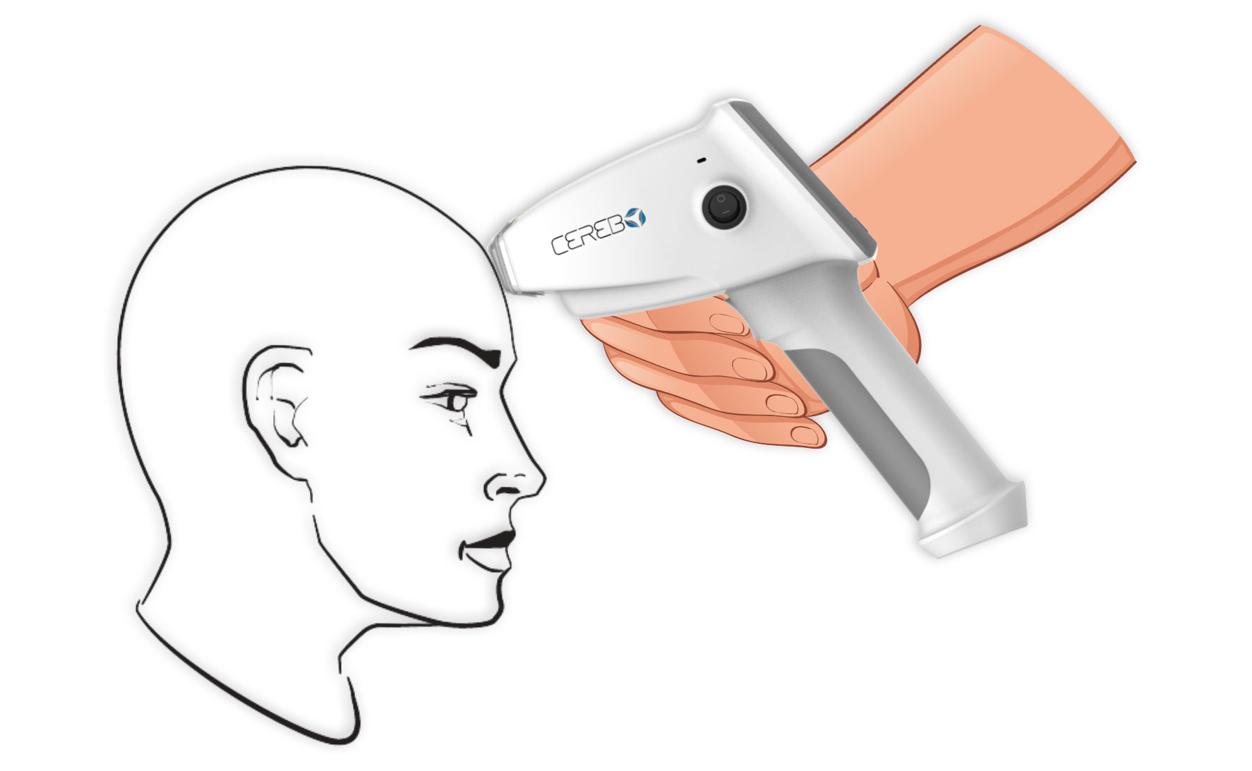
Early detection of traumatic brain injury
 Reference: BrainLine 2018, Managing Pain After Brain Injury, BrainLine, viewed on Apr 2020, Reference
 Reference: Dewan M-C 2020, Estimating the global incidence of traumatic brain injury, Journal of Neurosurgery, viewed on Apr 2020, Reference
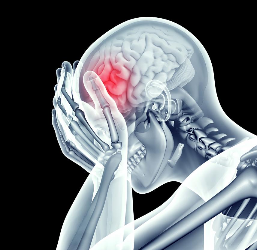
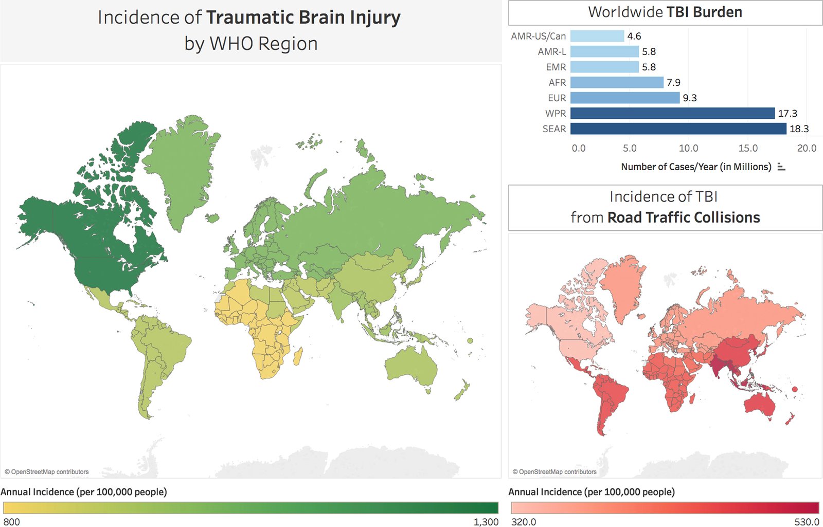
More than 50 million suffer from traumatic brain injury each year and 50%-90% of patients with mild traumatic brain injury go unidentified or undiagnosed[1,2]. TBI is never left untreated intentionally. Unfortunately, it is hard to diagnose symptoms in the beginning and know what symptoms the patients may suffer. Also symptoms of traumatic brain injury can be misleading due to overlap with those of other disorders[3].This leads to delayed detection and hence delayed treatment. For this reason, traumatic brain injury is associated with a very high mortality and disability rate. WHO predicts that TBI will be the leading cause of disability in the world by 2020.
Reference:
[1] CENTER-TBI 2020, Traumatic Brain Injury Fact sheets and Policy brief, European Brain Injury Consortium, viewed 09 Apr 2020, Reference
[2] Prince C, Bruhn M-E 2017, ‘Evaluation and Treatment of Mild Traumatic Brain Injury: The Role of Neuropsychology’, Brain Sciences, 7(8), pp 105, viewed 30 Apr 2020, Reference
[3] Brain Injury Law center 2020, UNTREATED TBI: WHAT ARE THE EFFECTS OF AN UNTREATED CONCUSSION?, viewed 09 Apr 2020, Reference
Deep brain spectroscopy to investigate the brain tissue in-vivo
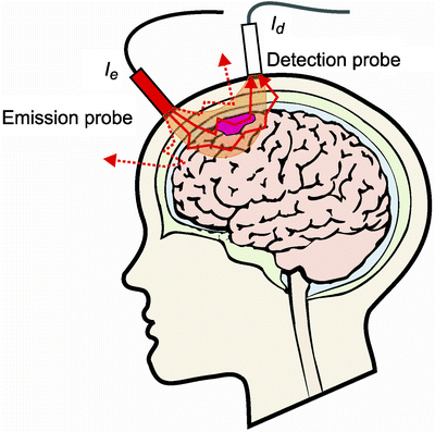
The optical methods used for neuromonitoring are based on emission of near-infrared light (NIR) at the surface of the head and detection of remitted light at a distance of several centimeters. Two main phenomena associated with light are: scattering and absorption. As described by diffusion theory, Scattering depends on the cellular structure of the tissue and leads to stochastic movement of photons in the medium. The strong scattering and good transparency of tissue layers of the head for NIR light result in sufficient re-emission of photons back to the surface to allow for detection of photons that penetrated the brain cortex. The chromophores found in the tissue like hemoglobin, water, lipids, and a variety of proteins (such as cytochrome c) have varied light absorption spectra.
CEREBO®'s proprietary optoelectronic configuration deploying diffuse reflectance spectroscopy detects the presence of intracranial hemorrhage non invasively
 Reference: Okada E 2013, Photon Migration in NIRS Brain Imaging, Application of Near Infrared Spectroscopy in Biomedicine, Vol 4, pp 37-58, viewed Apr 2020, Reference

NIRS

ML Algorithms

Software

Hardware
CEREBO®'s novel application of near infrared spectroscopy makes it possible to detect intracranial hemorrhage non invasively. Correlation of optical density of near infrared light and hemoglobin concentration is well established in science
Machine Learning powered algorithms eliminates the need of expert data interpretation. Hence Cerebo becomes usable by a common man with minimal training.
This device becomes very easy to use by the virtue of patented zero calibration system and intuitive interface.
The miniaturized hardware is designed to bridge the limitations of traditional tools that are bulky, expensive, and need much greater infrastructure support like air conditioning to provide point-of-care detection that will assist in triaging suspected TBI patients in military, sports, and emergency or urgent care environments both in India and internationally.
Generates result for one patient
at a time
Maintains history for 100 most recent patients
All the patients data available on cloud accessible via app and web application